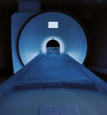
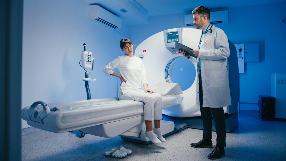
An MRI can give your healthcare provider a closer look at an injury or a suspected medical condition. The information it provides depends on the body area being examined and the reason for the scan.
A lumbar MRI focuses on the lower spine, while brain, knee and shoulder MRI examinations answer different questions about those structures.
At Advanced Diagnostic Radiology in Cumberland, Maryland, we provide diagnostic imaging to support your provider’s evaluation and treatment planning.
Here is what these four types of MRI may reveal, when they may be recommended and what to discuss before scheduling.
How MRI Helps Your Provider Evaluate Symptoms
Magnetic resonance imaging uses a strong magnetic field, radio waves and a computer to create detailed images without ionizing radiation. It is especially useful for examining structures such as spinal discs, nerves, ligaments, tendons and brain tissue.
Your provider selects the examination based on your symptoms, medical history and physical examination. An MRI is not automatically the first test for pain, and the images do not always explain every symptom. Findings must be interpreted in context. Our MRI service page offers an overview of MRI at ADR.
Back MRI for Lower Back Pain and Nerve Symptoms
A lumbar MRI examines the lower portion of your spine, including the vertebrae, discs, spinal canal and nearby nerves.
Your provider may consider it when back pain persists despite recommended treatment, when symptoms suggest nerve involvement or when the examination raises concern about a more serious condition.
Pain traveling into the buttocks or legs, numbness, tingling or weakness may prompt further assessment. The decision to order imaging depends on the full clinical picture, rather than the presence of any single symptom.
As Cleveland Clinic’s lower back pain guide explains, back pain has several possible causes and often improves with nonsurgical care.
A lumbar MRI may help evaluate:
- Bulging or herniated discs and their relationship to nearby nerves.
- Spinal stenosis, or narrowing of spaces within the spine.
- Selected injuries involving vertebrae, ligaments or other spinal structures.
- Suspected infection, inflammation or tumors when clinically indicated.
RadiologyInfo’s spine MRI guide describes how the examination can assess discs, nerves and other spinal tissues.
Some imaging changes may be unrelated to the pain you are experiencing, so your provider will compare the scan with your examination before recommending treatment.
Brain MRI for Neurological Symptoms
A brain MRI, also called a head MRI, creates detailed pictures of brain structures.
A provider may recommend it to investigate selected neurological symptoms, such as persistent or changing headaches, unexplained dizziness, seizures or changes in vision, depending on the history and examination. Not every headache requires an MRI.
Depending on the protocol, a brain MRI may help evaluate:
- Brain tumors and other structural abnormalities.
- Stroke-related injury and certain blood vessel abnormalities, sometimes requiring specialized imaging sequences.
- Lesions associated with multiple sclerosis.
- Selected infections, inflammation or changes in brain tissue.
Mayo Clinic provides useful background on brain tumors and multiple sclerosis. These are among the conditions a provider may investigate; being referred for MRI does not mean you have one of them.
The examination may be ordered with or without contrast. Specialized scans may be needed to assess blood vessels or answer other focused questions.
A brain MRI overview from RadiologyInfo explains common uses and how a radiologist interprets the images.
Knee MRI for Pain Swelling or an Injury
A knee MRI can examine the soft tissues, cartilage and bone around the joint. Your provider may consider it when symptoms or an injury suggest damage that needs more detail than an X-ray can provide. Examples include persistent swelling, instability, reduced motion or symptoms after a sports injury.
The scan can help assess ACL and PCL injuries, meniscus tears, tendon injuries and cartilage damage. It may also reveal bone bruising or certain fractures that are difficult to see on other tests. RadiologyInfo’s knee MRI guide outlines these applications.
The menisci are cartilage structures that help cushion the knee. A tear can be relevant to symptoms, but the scan alone does not determine whether surgery is needed. Your provider considers the location and extent of the finding, your examination and how symptoms affect your activities before discussing treatment options.
Shoulder MRI for Persistent Pain or Limited Movement
A shoulder MRI provides detailed images of the rotator cuff, tendons, ligaments, cartilage and surrounding structures. Your provider may recommend it for pain that has not improved with initial care, weakness after an injury, recurrent instability or suspected soft-tissue damage.
The examination may help identify rotator cuff tears, biceps tendon injuries, bursitis and damage to the shoulder labrum, the rim of tissue that helps stabilize the joint. It can also evaluate selected bone abnormalities. RadiologyInfo’s shoulder MRI guide explains these uses and the role of specialized examinations.
For some clinical questions, your provider may request an MR arthrogram, which involves injecting contrast into the joint before MRI. This differs from a routine shoulder MRI or an MRI using IV contrast. Confirm the exact examination on your order when scheduling.
Preparing for Your Brain Back Knee or Shoulder MRI
Preparation is similar for many MRI examinations, but your instructions depend on the scan and whether contrast or sedation is planned. Before your appointment:
Tell the team about implants, pacemakers, surgical clips, stimulators or metal fragments. Provide device identification information so the exact model and scanning conditions can be reviewed.
Mention kidney problems, previous contrast reactions, recent surgery and any possibility of pregnancy.
Follow the specific eating, drinking and medication instructions supplied for your examination.
Discuss claustrophobia or difficulty lying still in advance. If a sedative is prescribed, arrange a driver and follow the discharge instructions.
Ask whether to bring your imaging order, prior images and insurance information. Remove metal items and change into a gown as directed.
During the examination, you will lie on a movable table while the technologist positions the body area being scanned. The machine makes loud tapping or knocking sounds, so hearing protection is provided. Remaining still helps the team obtain clear images, and the technologist can communicate with you throughout the scan.
Many MRI scans take approximately 20 to 60 minutes, although some take longer. Allow additional time for check-in, safety screening and any IV preparation. Ask for an estimate for the examination your provider ordered.
Understanding Your MRI Results
A radiologist reviews the images and sends a report to your referring healthcare provider. Your provider then explains the findings alongside your symptoms and medical history.
Depending on the results, next steps may include continued conservative care, a specialist consultation or additional testing.
Ask when the report is expected and which office will contact you. Results are not usually interpreted for you immediately after the scan, and timing varies. An MRI finding does not automatically establish the cause of a symptom or mean that surgery is necessary.
Planning an ADR Visit From Leesburg Baltimore Hagerstown or Martinsburg
If you are researching a back MRI from Leesburg, VA, a brain MRI from Baltimore, MD, a knee MRI from Hagerstown, MD, or a shoulder MRI from Martinsburg, WV, confirm the imaging address and examination availability before booking travel.
ADR is located at 952 Seton Drive, Suite 210, Cumberland, MD 21502. Contact our team to confirm the ordered scan, scheduling options and preparation requirements.
Review our insurance and billing information and ask your insurer about network status, prior authorization and expected out-of-pocket costs.
Frequently Asked Questions
Is MRI always needed for back knee or shoulder pain?
No. Your provider may begin with an examination, conservative care or another imaging test. MRI is ordered when the additional detail is likely to help answer a clinical question.
Can one MRI examine all four areas?
These are separate targeted examinations with different positioning and imaging protocols. Ask which body area your order covers; additional areas require separate clinical consideration and scheduling.
Does every MRI require contrast?
No. Contrast depends on the clinical question. A routine joint MRI, an MRI with IV contrast and an MR arthrogram are different examinations, so confirm your specific order.
Does an abnormal MRI mean I need surgery?
No. Some findings can be managed without surgery, and others may not explain your symptoms. Treatment decisions depend on your examination, symptoms, goals and the nature of the finding.
Schedule Your MRI at Advanced Diagnostic Radiology
If your provider has recommended a brain, back, knee or shoulder MRI, call 301-777-3522 or contact ADR to discuss the next steps.
Our team can confirm appointment details and preparation instructions for your visit to our Cumberland office.