Tremor, stiffness, slowed movement, balance changes and walking difficulties can raise questions about Parkinson’s disease, but these symptoms can have several causes.
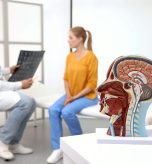
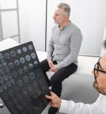
A neurological examination and medical history remain the foundation of diagnosis. Brain imaging may provide additional information when the cause is uncertain or a physician needs to evaluate other possible conditions.
Understanding brain scans for Parkinson’s disease can help patients see why a doctor may recommend DaTscan, PET or MRI.
These studies are not interchangeable: each examines a different aspect of the brain, and no scan should be treated as a stand-alone confirmation of Parkinson’s disease.
Early Signs That May Lead to an Evaluation
Parkinson’s symptoms often develop gradually. Possible early changes include:
- A tremor that is more noticeable at rest
- Slower movement or difficulty starting movement
- Muscle stiffness
- Changes in walking, posture, balance or coordination
- Smaller handwriting
- Changes in speech or facial expression
Having one of these symptoms does not mean a person has Parkinson’s disease. Essential tremor, medication effects, metabolic conditions and other neurological disorders can produce overlapping signs.
The first step is an evaluation by a qualified physician, who can decide whether imaging is likely to add useful information.
Can a Brain Scan Diagnose Parkinsons Disease
No single brain scan confirms Parkinson’s disease by itself. The diagnosis is generally based on the pattern and progression of symptoms, medical history and neurological findings.
Imaging may support that evaluation, help distinguish certain movement disorders or identify structural explanations for symptoms.
A normal or abnormal scan must be interpreted in context. The neurologist or referring physician considers what the scan measures, the patient’s examination and whether the result changes the diagnostic plan.
How DaTscan Supports Parkinsons Evaluation
DaTscan is a specialized nuclear medicine study performed with single-photon emission computed tomography, or SPECT. After a small amount of radiotracer is administered and allowed time to circulate, a gamma camera creates images of dopamine transporter activity in the brain.
DaTscan may help when a physician is trying to determine whether unclear movement symptoms are associated with reduced dopamine transporter activity. For example, it can support the distinction between a neurodegenerative parkinsonian syndrome and essential tremor when the clinical findings overlap.
An abnormal DaTscan is not specific to Parkinson’s disease. Other degenerative parkinsonian disorders may also produce reduced activity, so the study usually cannot identify the exact disorder responsible for an abnormal result.
What a Brain PET Scan Can Show
Positron emission tomography, or PET, uses a radiotracer, specialized camera and computer to evaluate activity or function in tissues. In brain imaging, the information available depends on the tracer selected and the clinical question being investigated.
PET may be considered when a physician needs tracer-specific information about brain metabolism, function or another neurological process. It does not provide the same information as DaTscan, and one study is not automatically a substitute for the other.
Why a Physician May Order Brain MRI
MRI uses a powerful magnetic field and radio waves to create detailed images of brain structure without ionizing radiation. It does not directly measure dopamine transporter activity, but it may help physicians look for structural abnormalities or other causes of movement-related symptoms.
For some patients, MRI is part of the broader evaluation rather than a test for Parkinson’s disease itself. The choice between MRI, DaTscan, PET or another study depends on the symptoms and the specific question the referring physician needs answered.
DaTscan vs. PET vs MRI for Parkinsons Symptoms
- DaTscan uses SPECT imaging to evaluate dopamine transporter activity.
- Brain PET evaluates tracer-specific activity, metabolism or function.
- Brain MRI provides detailed structural images and may reveal other explanations for symptoms.
- None of these studies should be interpreted without the patient’s clinical evaluation.
What to Expect From Advanced Brain Imaging
Preparation and appointment length depend on the ordered study. DaTscan and PET involve administration of a radiotracer followed by a waiting period and imaging. MRI requires patients to remove metal items and notify the facility about implants, devices or metal in the body.
Provide the imaging team with a complete list of medications, supplements, allergies, recent illnesses and relevant medical conditions. Some medications may interfere with specific nuclear medicine studies, but do not stop taking a medication unless the referring physician or imaging team instructs you to do so.
Patients who are pregnant, may be pregnant or are breastfeeding should notify the care team before imaging.
Brain Imaging Near Cumberland and Hagerstown, Maryland
Advanced Diagnostic Radiology provides MRI, PET and nuclear medicine imaging at 952 Seton Drive, Suite 210, in Cumberland, Maryland. The center also serves patients seeking advanced brain imaging near Hagerstown and throughout Western Maryland, as well as nearby communities in Pennsylvania and West Virginia.
If your physician has recommended imaging for Parkinson’s symptoms or another neurological concern, contact ADR to confirm availability, referral requirements and preparation instructions for the specific study.
Call Advanced Diagnostic Radiology at 301-777-3522 or request an appointment online. Your referring physician can explain why the examination was ordered and how the results fit into your overall evaluation.